
Morpho-molecular spatial profiling of colorectal cancer and the tumor budding niche
Aim
Tumor budding, single cancer cells or clusters of up to four cells at the invasive front, is one of the strongest histological indicators of metastatic risk in colorectal cancer, yet what drives it on the molecular level remains poorly understood, mainly because bulk sequencing mixes buds together with the surrounding stroma and immune cells. This project is built on spatial transcriptomics, which measures gene expression while preserving tissue architecture. CRC resections and pre-treatment rectal biopsies are profiled with the GeoMx, which measures the whole transcriptome separately in the cancer cell compartment and the tumor microenvironment, while CosMx adds true single-cell resolution in high-risk stage II patients, the group in which the decision whether to give chemotherapy is hardest. On the same tissues we run SpatioMorph, an image analysis pipeline we developed that segments individual cells and computes several hundred interpretable morphological features, so that what a tumor looks like can be linked directly to the molecular programs underneath. We describe colorectal cancer, and tumor budding in particular, on three complementary levels, morphological, molecular and morpho-molecular, guided by the questions of how metastatic dissemination via tumor budding occurs on the cellular and molecular level, which biomarkers could improve patient stratification beyond TNM staging, and which markers could predict therapy response in rectal cancer.
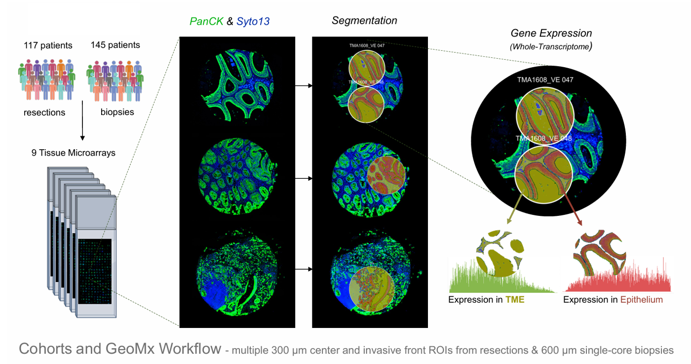
Fig 1. Cohorts and GeoMx workflow: multiple 300 µm center and invasive front ROIs from resections and 600 µm singlecore biopsies, stained for PanCK and Syto13, segmented into cancer and microenvironment compartments and profiled for the whole transcriptome in each.
Members

Jacob Hanimann

Inti Zlobec
Funding source

Hannah Williams