
Morphological profiling of lymph node metastases to refine prognosis in Stage III colorectal cancer
Aim
In colorectal cancer, whether the tumor has spread to the lymph nodes defines Stage III disease and determines adjuvant chemotherapy. But Stage III is not uniform, as patients with the same nodal stage can follow very different courses. Part of the reason may be that TNM staging counts how many nodes are involved without describing what the metastases actually look like, even though nodal metastases are thought to be one of the gateways to distant spread. We therefore ask: does the morphology of the metastatic deposit itself carry prognostic information beyond the node count?
To find out, we built and validated a lymph-node and metastasis segmentation pipeline (MetAssist 2.0 [1]) to automatically delineate lymph nodes and the tumor growing within them on H&E-stained whole slide images. From these regions we extract intra-metastatic morphological features grouped by theme — for example tumor budding, or the branching complexity of the metastatic glands — and summarize them from the level of individual lymph nodes up to the patient. After filtering out redundant and unstable features, we test them against patient outcome in survival models adjusted for standard clinicopathological factors, and combine the most informative into a single morphological signature. The aim is to obtain richer, quantitative readout of metastatic biology that could sharpen prognosis within Stage III.
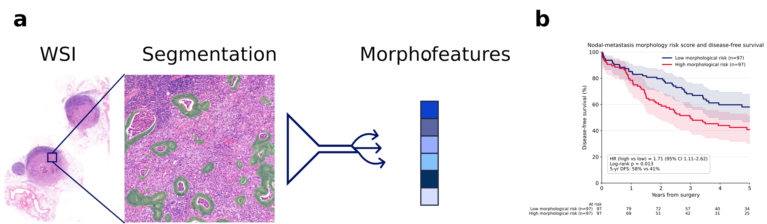
Figure 1. a) LN WSIs are first processed by a segmentation model that delineates the individual lymph-node fragments. A second segmentation model then outlines the metastatic tumour within them. Hand-crafted, interpretable morphological features are extracted from the resulting tumour mask, aggregated to the patient level, and used for survival analysis. b) Kaplan-Meier DFS by median split of the cross-validated morphology risk score. High vs low-risk groups: five-year DFS 41% vs 58%; HR 1.71 (95% CI 1.11–2.62); log-rank p = 0.013.
Members

Javier García Baroja

Inti Zlobec

Amjad Khan
Funding source
Collaboration
Iris Nagtegaal (Radboudumc)
Robert Zboray (EMPA)
Henning Müller (HES-SO)
Ludovico Silvestri (University of Florence)
Publications
[1] Garcia-Baroja, J., Dislich, B., Zens, P., Zagrapan, B., Parokkaran, F. M., Wütschert, L., Weber, S. E., Christe, L., Neppl, C., Rau, T., Perren, A., Tolkach, Y., Zlobec, I. & Khan, A. MetAssist 2.0: a generalizable AI framework for lymph node metastasis detection across multiple cancer types. Modern Pathology 39(6), 101003 (2026). DOI