
Three-dimensional characterization of colorectal cancer tissue by light-sheet microscopy
Aim
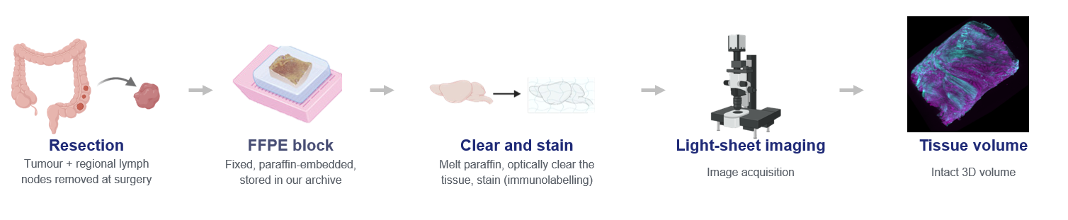
Diagnostic pathology examines tissue through thin sections, so every structure is judged from a single arbitrary plane. Features that are genuinely three-dimensional - the shape of the tumor invasion front, whether a tumor bud is truly detached from the main tumor body, how stroma and extracellular matrix are organized around it - are therefore only partly accessible, and some appearances are artifacts of how the block was cut. In this project, intact tissue punches from colorectal cancer resections are cleared and imaged by light-sheet fluorescence microscopy, producing whole volumes with nuclear, collagen and pan-cytokeratin channels at cellular resolution. Deep learning methods are used to segment the cytokeratin-positive tumor compartment, individual nuclei and the surrounding stromal architecture. From these segmentations, the geometry of the tumor surface is quantified through curvature, tortuosity and the connectivity of protruding structures; candidate tumor buds are traced through the volume to establish whether they are isolated or attached; and the composition and spatial organization of the microenvironment are compared across regions of differing surface morphology. Re-slicing the volumes into virtual 2D sections allows these measurements to be related directly to conventional histopathological assessment.
Members

Inti Zlobec
Funding source
Collaboration
Ludovico Silvestri (University of Florence)
Loredana Daminescu (ITMP, University of Bern)

Ekaterina Sedykh

Amjad Khan

Hannah Williams