
Tumor topology to predict outcome after neoadjuvant treatment in rectal cancer
Aim
Patients diagnosed with locally advanced rectal cancer are treated with neoadjuvant chemoradiotherapy to shrink the tumor before its resection and reduce the risk of local recurrence. Once the tumor is removed, pathologists assess how well it responded to the treatment using tumor regression gradings (TRGs), which estimate the amount of tumor remaining in the tissue. However, these gradings are difficult to reproduce and often correlate poorly with how patients actually fare, especially for the many patients showing an intermediate response. There is therefore a need for more reliable ways to read the response to treatment directly from the histology slides.
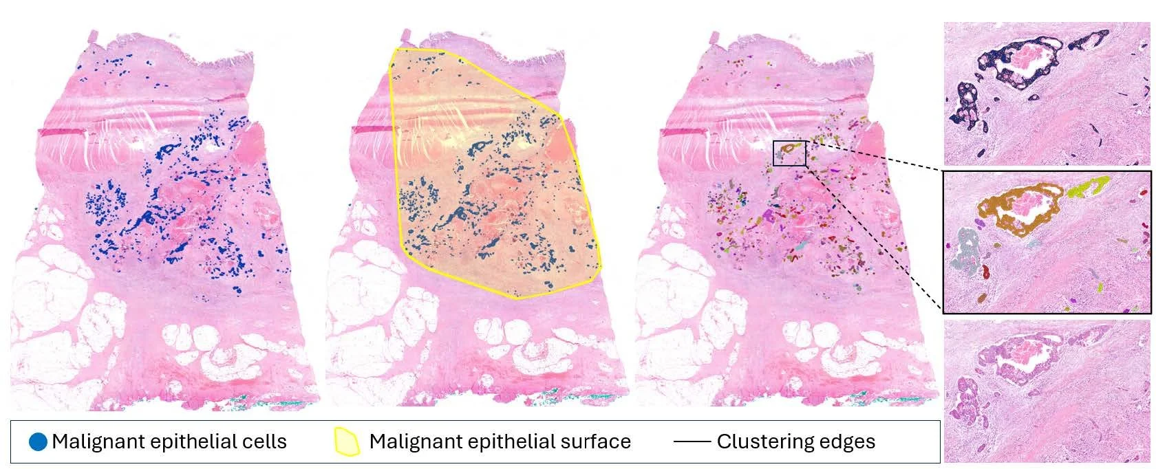
In this project, we aim at exploring the architecture of the tumor that remains after neoadjuvant treatment. We use deep learning models to detect and locate every residual malignant cell across the resection whole slide images, and group neighbouring cells together to reconstruct the individual tumor masses left in the tissue. From this we describe the tumor topology, the area the tumor covers and the size of its remaining cell clusters, and use it to propose a new patient stratification. We compare this stratification to the standard tumor regression gradings and evaluate its potential to guide patient follow-up, helping to tell apart patients who could be safely monitored from those who might benefit from additional treatment.

Members

Ana Leni Frei
Inti Zlobec

Andreas Fischer
Funding source
Collaboration

Sergio Vazquez Montes de Oca